Dr Damian Muñoz

Dr Damian Muñoz
Curso mínima invasiva south beach

domingo, 29 de diciembre de 2013

Ciatalgia


lunes, 23 de diciembre de 2013

The spine

FUENTE:
http://www.nature.com/nature/journal/v503/n7475_supp/full/503S1a.html#/author-information


The spine

Nature
 
503,
 
S1
 
 
doi:10.1038/503S1a
Published online
 

Every year up to a quarter of a million people around the world suffer a spinal-cord injury. These devastating injuries can end lives, rob people of their mobility and burden healthcare systems. As this Outlook shows, advances in medicine and technology are offering new ways to reduce pain and restore mobility.
Researchers are developing a variety of approaches to repairing spinal injuries. Some techniques use stem cells or reprogram existing cells to help the body recreate neurons damaged in an injury (page S4). Regenerative techniques are already helping to mend vertebrae, and replacement discs are being developed that closely mimic their natural components (S7). New drugs can treat pain, improve the level of recovery after an injury, and possibly stimulate biological mechanisms to replace damaged cells (S10). And biomechanical engineer Peter Cripton shows how collecting neck-injury data can trigger ideas for designing better safety devices, such as helmets that protect the head and neck (S13). A growing body of data shows that the consequences of such an injury depend critically on the emergency treatment provided, including medications that limit the damage (S14). If medical action is not enough, the latest technologies could help. Mechanical systems called exoskeletons can help a paraplegic to stand, walk and even climb stairs (S16).
For spine injuries, as with all healthcare, prevention is better than cure. Rehabilitation and prevention specialist Sara Klaas argues that safer behaviour, from avoiding multitasking when driving to eliminating trip hazards, can reduce the chances of an injury and save billions of dollars (S18). Ultimately, a combination of treatments from immediately after an injury to decades beyond can turn a devastated patient into a productive one with a fulfilling life.
We acknowledge the financial support of Mesoblast in producing this Outlook. As always, Naturehas full responsibility for all editorial content.

Author information

Affiliations

  1. Contributing Editor

    • Mike May

jueves, 12 de diciembre de 2013

Lumbar Compression Fracture

http://emedicine.medscape.com/article/309615-overview#showall

Contributor Information and Disclosures
Author
Andrew L Sherman, MD, MS  Associate Professor of Clinical Rehabilitation Medicine, Vice Chairman, Chief of Spine and Musculoskeletal Services, Program Director, SCI Fellowship and PMR Residency Programs, Department of Rehabilitation Medicine, University of Miami, Leonard A Miller School of Medicine

Andrew L Sherman, MD, MS is a member of the following medical societies: American Academy of Physical Medicine and RehabilitationAmerican Association of Neuromuscular and Electrodiagnostic MedicineAmerican Medical Association, and Association of Academic Physiatrists

Disclosure: Pfizer Honoraria Speaking and teaching
Coauthor(s)
Nizam Razack, MD  FACS, JD, Assistant Professor of Neurological Surgery, Orthopedics, and Rehabilitation, University of Central Florida Medical School; Neurosurgeon, Spine and Brain Neurosurgery Center; Chairman, Department of Neurosurgery, Orlando Regional Medical Center

Nizam Razack, MD is a member of the following medical societies: American Association of Neurological SurgeonsAmerican College of SurgeonsCongress of Neurological SurgeonsFlorida Medical Association, and Society for Neuro-Oncology

Disclosure: Nothing to disclose.
Specialty Editor Board
Curtis W Slipman, MD  Director, University of Pennsylvania Spine Center; Associate Professor, Department of Physical Medicine and Rehabilitation, University of Pennsylvania Medical Center

Curtis W Slipman, MD is a member of the following medical societies: American Academy of Physical Medicine and RehabilitationAssociation of Academic PhysiatristsInternational Association for the Study of Pain, and North American Spine Society

Disclosure: Nothing to disclose.
Francisco Talavera, PharmD, PhD  Adjunct Assistant Professor, University of Nebraska Medical Center College of Pharmacy; Editor-in-Chief, Medscape Drug Reference

Disclosure: Medscape Salary Employment
Patrick M Foye, MD  Associate Professor of Physical Medicine and Rehabilitation, Co-Director of Musculoskeletal Fellowship, Co-Director of Back Pain Clinic, Director of Coccyx Pain Service (Tailbone Pain Service: www.TailboneDoctor.com), University of Medicine and Dentistry of New Jersey, New Jersey Medical School

Patrick M Foye, MD is a member of the following medical societies: American Academy of Physical Medicine and RehabilitationAmerican Association of Neuromuscular and Electrodiagnostic Medicine,Association of Academic Physiatrists, and International Spine Intervention Society

Disclosure: Nothing to disclose.
Kelly L Allen, MD  Medical Director, Medevals

Disclosure: Nothing to disclose.
Chief Editor
Rene Cailliet, MD  Professor-Chairman Emeritus, Department of Rehabilitation Medicine, University of Southern California School of Medicine; Former Director, Department of Rehabilitation Medicine, Santa Monica Hospital Medical Center

Rene Cailliet, MD is a member of the following medical societies: American Academy of Pain Medicine,American Academy of Physical Medicine and RehabilitationAmerican Pain SocietyAssociation of American Medical CollegesInternational Association for the Study of Pain, and Pan American Medical Association

Disclosure: Nothing to disclose.


Background

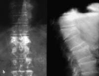
The lumbar vertebrae are the 5 largest and strongest of all vertebrae in the spine. These vertebrae comprise the lower back. They begin at the start of the lumbar curve (ie, the thoracolumbar junction) and extend to the sacrum. The strongest stabilizing muscles of the spine attach to the lumbar vertebrae. Fractures of lumbar vertebrae, therefore, occur in the setting of either severe trauma or pathologic weakening of the bone. Osteoporosis is the underlying cause of many lumbar fractures, especially in postmenopausal women. Osteoporotic spinal fractures are unique in that they may occur without apparent trauma. However, a thorough diagnostic workup is always required to rule out spinal malignancy. The image below reveals a wedge compression fracture. (See Pathophysiology.)
Anteroposterior and lateral radiographs of an L1 oAnteroposterior and lateral radiographs of an L1 osteoporotic wedge compression fracture.
In the past, treatment options for lumbar fractures were quite limited, with bracing and rest prescribed most often. While many patients improved with this regimen, some did not and were left with chronic, disabling pain. Suh and Lyles found that vertebral compression fractures were associated with significant performance impairments in physical, functional, and psychosocial domains in older women.[1]However, medical and surgical options are now available that can relieve the severe pain and disability from these fractures.

Recent studies

In a study of 55 patients with vertebral compression fracture, Rapan et al investigated changes in pain intensity following vertebroplasty (injection of a cement polymer into the fractured vertebral body; see Other Treatment). Treatment was administered to a total of 28 thoracic and 57 lumbar vertebrae; patients in the study had sustained vertebral fractures from spinal metastases or osteoporosis.
Prior to surgery, the patients' average pain score on the Visual Analog Scale (VAS) was 8.36, while 24 hours postsurgery it had fallen to an average of 2.23. At 3-month follow-up, the reduction in the VAS score remained nearly the same. Among the study's patients, 1 serious complication, paraparesis resulting from cement leakage into the spinal canal, occurred. The authors concluded that in patients with vertebral compression fracture who undergo vertebroplasty, the degree of pain reduction that occurs by 24 hours postsurgery predicts the intensity of pain patients will be experiencing 3 months later.[2]

Pathophysiology

The lumbar spine provides both stability and support, allowing humans to walk upright. Proper function of the lumbar spine requires that it have a normal posture (ie, a normal lumbar curve). Any injury that changes the shape of a lumbar vertebra will alter the lumbar posture, increasing or decreasing the lumbar curve. As the body attempts to compensate for the alteration in the lumbar spine in order to maintain an upright posture, this will tend to distort the curves of the thoracic and cervical spine.
Lumbar compression fractures can be a devastating injury, therefore, for 2 reasons. First, the fracture itself can cause significant pain, and this pain sometimes does not resolve. Second, the fracture can alter the mechanics of the posture. Most often, the result is an increase in thoracic kyphosis, sometimes to the point that the patient cannot stand upright. In trying to maintain their ability to walk, patients with kyphosis report secondary pain in their hips, sacroiliac joints, and spinal joints. These patients are also at risk for falls and accidents, increasing the risk of secondary fractures in the spine and elsewhere.
Fractures in the lumbar spine occur for a number of reasons. In younger patients, fractures are usually due to violent trauma. Car accidents frequently cause flexion and flexion distraction injuries. Jumps or falls from heights cause burst fractures. These fractures can also result in serious neurological injury. In older patients, lumbar compression fractures usually occur in the absence of trauma, or in the context of minor trauma, such as a fall. The most common underlying reason for these fractures in geriatric patients, especially women, is osteoporosis. Other disorders that can contribute to the occurrence of compression fractures include malignancy, infections, and renal disease.

Traumatic fractures

Different types of fractures can occur in the lumbar (or thoracic) spine. Classification of these fractures is based on the 3-column anatomic theory of Denis, which describes anterior, middle, and posterior spinal columns consisting of aspects of the spine and their corresponding ligaments and other soft-tissue elements. The Denis system, however, was created to classify traumatic fractures. A similar classification system does not exist for compression fractures. The main reason to use such a classification is to help determine whether a fracture is stable. Instability in the Denis system implies that damage has occurred to at least 2 of the columns of the lumbar spine.
  • Wedge fractures are the most common type of lumbar fracture and are the typical compression fracture of malignancy or osteoporosis. They occur as a result of an axially directed central compressive force combined with an eccentric compressive force. In pure flexion-compression injuries, the middle column remains intact and acts as a hinge. Although wedge fractures are usually symmetric, 8-14% are asymmetric and are termed lateral wedge fractures.
  • Fractures involving flexion and distraction forces are often due to lap belts in motor vehicle accidents. Commonly, the posterior columns are compromised in these injuries because the ligaments of the posterior elements are disrupted. This type of injury is quite common in young children. Most patients with flexion-distraction injuries remain neurologically intact.
  • Burst fractures result from high-energy axial loads to the spine. Multiple classification systems exist for these fractures. The severity of the deformity, the severity of canal compromise, the extent of loss of vertebral body height, and the degree of neurologic deficit affect the determination of whether these injuries are unstable.
When any of the above injuries occurs with a severe rotational force, the degree of injury and of instability increases.

Nontraumatic fractures

In osteoporosis, osteoclastic activity exceeds osteoblastic activity, resulting in a generalized decrease in bone density. The osteoporosis weakens the bone to the point that even a minor fall on the tailbone, causing an axial load or flexion, results in one or more compression fractures. The fracture is usually wedge shaped. Without correction, a wedge fracture invariably increases the degree of kyphosis.
Malignancies that result in spinal fractures are most commonly metastases rather than primary bone cancers. Primary cancers that often spread to the spine via hematologic dissemination include cancers of the prostate, kidneys, breasts, and lungs. Melanoma is a less common but more aggressive cause of spinal metastasis. The most common primary cancer of the spine is multiple myeloma, but others, including a variety of sarcomas,[3] can also manifest as a spinal fracture. Nonmalignant lesions that can cause fractures include aneurysmal bone cyst and hemangioma.
Spinal infections usually start in the lumbar intervertebral disk. From the disk, the infection spreads to bone, resulting in osteomyelitis. Severe pain is the hallmark symptom. The exception is spinal tuberculosis or Pott disease. In this case, the disk spaces are typically spared and a compression fracture may be the initial manifestation that leads to its discovery.

Epidemiology

Frequency

United States

Most fractures of the lumbar spine that require operative treatment occur at the thoracolumbar junction. These injuries are primarily traumatic in origin. Most nontraumatic lumbar fractures are osteoporotic in origin. These are almost invariably wedge-type compression fractures. The National Osteoporosis Foundation (NOF) estimates that currently, 10 million individuals in the United States have osteoporosis, and 34 million more have low bone mass.[4] In 2005, osteoporosis was responsible for more than 2 million fractures; approximately 547,000 of those were vertebral fractures. Approximately one third of osteoporotic vertebral injuries are lumbar, one third are thoracolumbar, and one third are thoracic in origin. Additionally, 75% of women older than 65 years who have scoliosis have at least 1 osteoporotic wedge fracture.

Mortality/Morbidity

  • Mortality from a lumbar fracture is rare; however, morbidity can be significant.
  • In elderly patients with acute osteoporotic fractures, pain and prolonged bed rest can lead to multiple secondary medical complications.
  • In younger persons, neurologic damage from traumatic spine injuries can result in problems such as loss of lower extremity strength and sensation and loss of bowel and bladder control.

Sex

Osteoporosis occurs primarily in postmenopausal women. Type 1 osteoporosis occurs in women aged 51-65 years and is associated with wrist and vertebral fractures. Estrogen deficiency is the main etiologic factor. Type 2 osteoporosis (senile type) is observed in women and men older than 75 years, in a 2:1 ratio of women to men.

Age

In young and middle-aged adults, most lumbar fractures are traumatic in origin. High-velocity falls can cause burst fractures, and seat-belt injuries can cause wedge fractures. As stated above, women 51-65 years old develop type 1 osteoporosis. After age 75 years, men also begin to develop type 2 osteoporosis.

History

Midline back pain is the hallmark symptom of lumbar compression fractures. The pain is axial, nonradiating, aching, or stabbing in quality and may be severe and disabling. The location of the pain corresponds to the fracture site, as seen on radiographs. In elderly patients with severe osteroporosis, however, there may be no pain at all as the fracture occurs spontaneously.
Young adults may present with severe back pain following an accident, such as a fall or a motor vehicle accident. Lower extremity weakness or numbness are important symptoms of neurologic injury from the fracture.
Vertebral fractures may also cause referred pain. Gibson et al presented a study of 350 patient encounters in 288 patients with 1 or more compression fracture without conus medullaris compromise or spinal nerve compression. They found that nonmidline pain was present in 240 of the 350 encounters. The pain was typically in the ribs, hip, groin, or buttocks. Treatment of the fracture with vertebroplasty (see Other Treatment) resulted in 83% of those patients gaining pain relief.[5]
Alternatively, many compression fractures are painless. Osteoporosis is a silently progressive disease. Osteoporotic compression fractures are often diagnosed when an elderly patient presents with symptoms such as progressive scoliosis or mechanical lower back pain and the physician obtains routine lumbar radiographs.
Finally, patients may present with a known (or unknown) malignancy. Routine spinal screening via magnetic resonance imaging (MRI; if focal or referred pain occurs), or via bone scan (as a survey if pain has not occurred) reveals the pathologic fracture. The most common malignancies leading to spinal involvement in the form of fractures are metastasis and multiple myeloma. Often, the compression fracture is the presenting manifestation that leads to the diagnosis of malignancy. However, patients may also have unexplained fevers, night sweats, past history of malignancy, or weight loss.
Finally, patients who have recently traveled outside of the United States, or who live in the inner city, may have symptoms of infection, such as general malaise, fever, or severely increasing pain. In these patients, osteomyelitis and Pott disease (tuberculosis spondylitis) must be ruled out.

Physical

A detailed neurologic examination is essential in all patients presenting with back pain, spine deformity, or traumatic spine injury. Most interventional procedures to alleviate pain in compression fractures are contraindicated in cases of neurologic compromise. Thus, a rectal examination is required to assess for rectal tone and sensation in trauma patients.
Upon inspection of the spine, the patient typically has a kyphotic posture that cannot be corrected. The kyphosis is caused by the wedge shape of the fractured vertebra; the fracture essentially turns the lateral conformation of the vertebra from a square to a triangle.
Hip flexor contractures due to iliopsoas shortening are typically present.
Palpation is important to correlate any reports of pain to the radiographic level of injury. Extreme pain elicited with superficial palpation is often observed in patients with spinal infections. Moderate pain is usually present at the level of the fracture.

Causes

The principal underlying cause of lumbar compression fractures is osteoporosis. In women, the leading risk factor for osteoporosis is menopause, or estrogen deficiency. Additional risk factors that may worsen the severity of osteoporosis include cigarette smoking, physical inactivity, use of prednisone and other medications, and poor nutrition. In males, all of the above nonhormonal risk factors apply; however, low testosterone levels also may be associated with compression fractures.
Renal failure and liver failure are both associated with osteopenia. Nutritional deficiencies can decrease bone remodeling and increase osteopenia. Finally, genetics also play a role in the development of compression fractures; osteoporosis can be observed in closely related family members.
Malignancy may manifest initially as a compression fracture. The most common malignancy in the spine is metastasis. Typical malignancies that metastasize to the spine are renal cell, prostate, breast, and lung, although other types can metastasize to the spine on rare occasions. The 2 most common primary spine malignancies are multiple myeloma and lymphoma.
Infection that results in osteomyelitis can also result in a compression fracture. Typically, the most common organisms in a chronic infection are staphylococci or streptococci. Tuberculosis can occur in the spine and is called Pott disease.


5 Tips for Flying Back Pain Free

http://www.spine-health.com/blog/5-tips-flying-back-pain-free


5 Tips for Flying Back Pain Free

By: Stephanie Burke

Flying with Back Pain
With the upcoming holidays, many people are starting to worry about how to manage the pain associated with a long flight.
Collected from many posts on our Forums, here are the top 5 most frequently mentioned tips about how to survive an airplane ride with less pain or discomfort.
  1. Move
    Sitting in the same position for a prolonged period puts a great deal of stress on your spine, so get up and walk and stretch as frequently as possible. Go to the back of the plane and do some gentle stretches - toe raises. Consider bringing a doctor's note and alert the flight crew prior to boarding that you have a back condition and will need to
  2. Schedule Smart
    Try to book a flight for a time of day when the plane is likely to be less full. With no one sitting next to you it will be easier to move and stretch while remaining in a sitting position, and to change sitting positions as needed. It will also be easier to retrieve your belongings from under the seat in front of you without twisting and straining your lower back.
  3. Support your Spine
    Bring a back roll or ask for extra pillows to put behind your back to keep your spine straight and prevent slouching. This will alleviate pain and pressure. If you are on the shorter side, bring something to prop up your feet to keep your knees at a right angle.
  4. Bring the Heat and Chill
    Bring gel packs that can be frozen or heated (or bring one of each). These are great for treating swelling, sore muscles, back pain, and even headaches. Be sure to have the physician's note about your back condition handy in case airport security has issues with the gel pack in your carry-on luggage. Bring a couple of empty Ziploc® baggies as well - these will not be an issue to get through security can be filled with ice once you're at the gate and/or on board the plane.
  5. Pain Medication can Help
    OTC pain killers like acetaminophen and NSAIDs, or prescription drugs like narcotics or muscle relaxants, can help "take the edge" off during and after the flight. Remember, these medications need to build up in your bloodstream, so it is best to take them at least an hour before you anticipate the pain setting in. Again, a letter from your doctor stating your need for any prescribed pain medications will help with possible airport security issues; and always be sure to keep medications in their original bottles.
Using a combination of the tips above should make travel as easy on your back as possible. And remember to try to reduce stress however possible - bring soothing music, a good book, chocolate, do some deep breathing - anything that eases your mind and calm your body will help.
lunes, 9 de diciembre de 2013

Burst Fracture C7

35 year fell on head now with neck pain. What's the diagnosis? 

Burst Fracture


· Burst fractures result from axial loading most often secondary to motor vehicle accidents and falls
· The axial load drives the intervertebral disk into vertebral body below
· Usually produces a comminuted, vertical fracture through the vertebral body
· Fragments may be retropulsed into the spinal canal injuring the cord
· Burst fractures may resemble flexion-teardrop fractures

o In a classical flexion-teardrop fracture, there is an avulsed anterior, inferior triangular bony fragment that is separated from the body and displaced anteriorly

o Both the anterior and posterior ligamentous structures are injured, which may not be the case in a burst fracture

§ Burst fractures, however, can have associated injury to the posterior ligamentous structures, especially if there is a combination of axial loading and flexion at the time of injury

· Clinically
o Neck pain

o Numbness or parasthesia

o Weakness

· Imaging on conventional radiography
o Lateral view of the cervical spine on conventional radiograph should show a comminuted fracture of the vertebral body

o Soft tissue swelling can be recognized by an increase in the prevertebral soft tissue of greater than ½ the AP diameter of the C3 vertebral body at C3 or greater than the full AP diameter of the cervical vertebral body at C6

o The anterior portion of the body will be wedged

o Retropulsion can be inferred if the posterior surface of the vertebral body is convex towards the spinal canal, as the normal cervical vertebral body has a concave posterior surface

o Injury to the posterior ligamentous structures can be inferred by widening of the interspinous distance and forward subluxation of the vertebral body above the fracture

o CT will show the comminuted fracture and the retropulsed fragment


Burst fracture, C7. Lateral view of the cervical spine demonstrates a comminuted vertical fracture
through the body of C7. The posterior surface of C7 is displaced posteriorly toward the spinal canal (red arrow)
while there is slight soft tissue swelling anteriorly (white arrow).
For a larger photo of the same image without the arrows, click here

· Treatment

o Burst fractures may be treated initially with cervical tongs

o The fracture is considered stable if there is no neurologic deficit or if there are no retropulsed fragments

miércoles, 4 de diciembre de 2013

Exam film


lunes, 2 de diciembre de 2013

OSTEOBLASTOMA VERETEBRA L4


FISIOMÓNICA: Efectos del Ejercicio Muscular Respiratorio- en la...

FISIOMÓNICA: Efectos del Ejercicio Muscular Respiratorio- en la...: Los ejercicios respiratorios musculares son utilizados no sólo en la rehabilitación de pacientes con enfermedades respiratorias, sino ta...


jueves, 28 de noviembre de 2013

La cirugía mínimamente invasiva de la columna vertebral cumple los objetivos

FUENTE:

http://neurocirugia.diariomedico.com/2010/11/05/area-cientifica/especialidades/neurocirugia/cirugia-minimamente-invasiva-de-columna-vertebral-cumple-objetivos


PABLO PALACIOS, DEL HOSPITAL UNIVERSITARIO MADRID SANCHINARRO

"La cirugía mínimamente invasiva de la columna vertebral cumple los objetivos"

"La técnica de cirugía mínimamente invasiva de la columna vertebral cumple todos los objetivos fijados, entre los que se encuentran la reducción de la agresión quirúrgica -con menor lesión de las partes blandas y menos sangrado y dolor postoperatorio- y el mantenimiento de los resultados de la cirugía abierta a medio y largo plazo".
A. C. M.   |  

Así lo ha indicado Pablo Palacios, del Hospital Universitario Madrid Sanchinarro, y uno de los directores de la Jornada Hispano-Lusa de Actualización en cirugía mínimamente invasiva y percutánea de la columna vertebral, celebrada en el citado centro.
Palacios ha presentado las conclusiones de un estudio, realizado en el Hospital Universitario Madrid Sanchinarro, que comparaba pacientes intervenidos con técnica convencional y otros a los que se les aplicó la cirugía mínimante invasiva, observando que no existen diferencias desde un punto de vista clínico a partir de los seis meses de la operación.
Por su parte, Juan Calatayud, neurocirujano del Hospital Clínico Universitario Lozano Blesa, de Zaragoza, ha resaltado que "la intención de los cirujanos de columna es mejorar y alargar la calidad de vida de un esqueleto con el que convivimos desde que nacemos hasta que morimos. Lo que queremos es que la gente viva mejor el mayor tiempo posible".
En la otra cara de la moneda se encuentran las limitaciones de esta técnica. "Tanto la curva de aprendizaje como el tiempo quirúrgico son mayores. Otra barrera de la cirugía mínimante invasiva es la mayor demanda tecnológica (fuentes de luz, lupas...)", ha indicado Palacios.
  • Desde el punto de vista clínico no existen diferencias entre la cirugía mínimamente invasiva y la abierta a partir de los seis meses de la operación
El representante portugués, Eurico Silva, de la Unidad Vértebro-Medular del Hospital General de San Antonio, en Oporto, ha añadido algunas limitaciones más: la obesidad, la osteoporosis, las deformidades como escoliosis y la espondilolistesis de alto grado. Sin embargo, ha añadido que las opciones de la cirugía mínimente son variadas. "Puede emplearse para la descompresión de estructuras neurológicas, la artrodesis y fijación vertebral y la vertebroplastia y cifoplastia".
Según Eduardo Hevia, cirujano de la columna vertebral del Hospital Universitario Madrid Sanchinarro, que ha codirigido el curso junto con Palacios, las complicaciones de la cirugía mínimamente invasiva percutánea son similares a las que puede haber con una técnica abierta. "Una posible complicación de este tipo es la malposición de los tornillos. Además, los errores en el manejo de la instrumentación son escasos pero peligrosos".
Por otro lado, Hevia ha destacado que las complicaciones de las indicaciones de la cirugía mínimamente invasiva percutánea son las más evitables. "Para ello hay que tener cuidado con las reintervenciones".
Otro de los ponentes, Antonio Mostaza, neurocirujano espinal del Hospital Universitario de León, tiene una amplia experiencia en cirugía mínimamente invasiva por endoscopia y defiende que con menos agresión se obtienen mejores resultados. Por ejemplo, la intervención de un paciente con hernia discal requiere una estancia postoperatoria de sólo unas horas ".

Unos 60 pacientes se beneficiaron de cirugía mínima invasiva de columna vertebral en Hospital Insular de Gran Canaria

FUENTE:
http://www.20minutos.es/noticia/615319/0/

Unos 60 pacientes se beneficiaron de cirugía mínima invasiva de columna vertebral en Hospital Insular de Gran Canaria

El Servicio de Neurocirugía del Hospital Universitario Insular de Gran Canaria ha realizado hasta el momento más de 60 intervenciones de cirugía con mínima invasión de la columna vertebral.

ECO ®  EUROPA PRESS. 26.01.2010 


Así, lo ha expuesto el jefe del Servicio de Neurocirugía del citado Hospital, Maximino González, y el médico adjunto del Servicio de Neurocirugía, Carlos Alberto Valencia, ambos han detallado los beneficios de esta técnica y las diferencias respecto a la intervención convencional, según informó el Ejecutivo regional en nota de prensa. La cirugía de mínima invasión sustituye a la cirugía abierta convencional en algunos casos. De todos modos, el Hospital Universitario Insular de Gran Canaria lleva realizando esta nueva técnica desde hace más de dos años. En concreto, dicha cirugía mínima invasiva de columna vertebral "necesita una menor" estancia hospitalaria tras la operación, ya que no es necesaria una transfusión sanguínea, se produce un "menor índice" de infecciones tras la intervención y se necesitan "menos" dosis de analgésicos. Además, la recuperación "es más rápida", produciendo "menos" dolor postoperatorio a largo plazo, mejorando el impacto estético y produciéndose un "menor daño" muscular. Así, la cirugía mínima invasiva se realiza en la columna vertebral cuando el paciente tiene patologías como la discopatía lumbar crónica, hernia discal, fracturas de columna con desviación y estenosis de canal de la zona lumbar. Actualmente, el Hospital Universitario Insular de Gran Canaria está realizando diferentes técnicas dentro de la cirugía mínima invasiva de columna vertebral, así como la implantación de tornillos vertebrales, dispositivos interespinosos, cifoplastía y vertebroplastía, implantación de dispositivo intradiscal, implantación por vía percutánea de prótesis que aumentan el calibre del canal vertebral e implantación de prótesis dentro de la vértebra. Para la intervención de este tipo de operaciones es necesaria la colaboración entre el Servicio de Neurocirugía, el Servicio de Anestesiología y el Servicio de Radiología del centro hospitalario.

Ver más en: http://www.20minutos.es/noticia/615319/0/#xtor=AD-15&xts=467263

Cirugía Mínimamente Invasiva

FUENTE:
http://ng-clinicas.com/especialidades/columna-2/cirugia-invasiva


El objetivo de la cirugía mínimamente invasiva es resolver patología con las mismas garantías de éxito que la cirugía tradicional, pero disminuyendo el daño causado por el acceso necesario al cuerpo. El concepto de MÍNIMA INVASIVIDAD puede llevar a engaño, ya que no se trata únicamente de disminuir el tamaño de las cicatrices. Si disminuyendo el tamaño de las cicatrices empeoramos la tasa de éxito, alargamos exageradamente la duración del procedimiento o no mejoramos el daño interno a las estructuras sanas, el proceso no es realmente poco invasivo, sino únicamente más estético. Por lo tanto, el término que mejor definiría a todas estas modernas técnicas de acceso quirúrgico es el de MÍNIMA AGRESIVIDAD.

La cirugía de columna ha sido pionera en la incorporación de técnicas mínimamente invasivas, mediante la aplicación de medios de visualización y el diseño de implantes específicos. Nuestro equipo profesional ha estado a la vanguardia en el uso de implantes percutáneos a nivel nacional. En 2003 el Dr. Ferrández practicó las primeras artrodesis MI-TLIF de España (técnica mínimamente invasiva), consiguiendo unas tasas de fusión idénticas a las del TLIF abierto, pero con una evidente reducción del tiempo de convalecencia y del daño muscular regional. Los primeros resultados se comunicaron en el V Congreso de Neuro-Raquis, en Gerona (2005).


Posteriormente se fueron introduciendo otras técnicas con implantes de mínima agresividad como la estabilización dinámica transpedicular (Dynesys) o interespinosa (X-Stop, DIAM, Aperius, etc). Hacia 2009 se puso en marcha la técnica de fijación trans-sacra AxiaLIF, que permite fusionar la unión lumbosacra a través de una única incisión de 2 cm a la altura del coxis. Y más tarde, en 2010, la fijación lumbar extremo-lateralXLIF.

Otro grupo de técnicas son las realizadas con agujas o trócares percutáneos, que permiten actuar sobre el disco intervertebral, las raíces nerviosas o los cuerpos vertebrales. Pueden infiltrarse sustancias tanto en el disco como alrededor de los nervios, introducir cemento en un cuerpo vertebral fracturado o bien utilizar sondas de radiofrecuencia para provocar la cicatrización retráctil del tejido discal (en caso de hernias contenidas) o la anulación selectiva de las fibras del dolor de alguna raíz nerviosa. Este tipo de técnicas se realizan de forma ambulatoria. La más novedosa de las técnicas intradiscales, disponible desde 2012, consiste en la reconstitución de la presión y composición de los discos degenerados, mediante la implantación de varillas de hidrogel GelStix. Los hidrogeles liberan sustancias correctoras del pH a la vez que aumentan más de 10 veces su volumen en contacto con la humedad de los tejidos, restaurando la altura y capacidad de amortiguación del disco.

Finalmente, las técnicas asistidas por endoscopia permiten actuar sobre el canal medular con un acceso mínimo al mismo, el cual evita daños a las estructuras sanas circundantes. La óptica VITOM permite realizar accesos a la columna torácica con pequeños abordajes retropleurales, evitando las secuelas propias de una gran toracotomía. Los microendoscopios de SpineView nos facilitan el acceso al canal lumbar, a través del neuroforamen, para la extracción de hernias discales bajo anestesia local y de forma ambulatoria en la mayor parte de los casos. Los foramenes neurales son agujeros a través de los cuales salen de la columna las raíces nerviosas lumbares para formar el nervio ciático en la pierna. Esos agujeros se convierten en la mejor vía natural de acceso al canal, evitando tener que abrirlo artificialmente.

Endoscopia Minimamente Invasivo en Columna Vertebral


Publicado el 01/03/2012
Dr. Jose Miguel Donoso especialista en dolores de la columna vertebral, trabaja en la Clinica Tabancura, Fundación Médica San Cristóbal y Clínica Avansalud.


miércoles, 27 de noviembre de 2013

Periprosthetic Patellar Fracture Fixation Using Suture Anchors

FUENTE:
http://www.healio.com/orthopedics/journals/ortho/%7B321eca22-84c7-4f40-99d9-bfd0099124fd%7D/periprosthetic-patellar-fracture-fixation-using-suture-anchors


CASE REPORT 

Periprosthetic Patellar Fracture Fixation Using Suture Anchors

Rajesh N. Maniar, MS(Orth), MCh Orth, FCPS, DNB; Ravi M. Nayak, MS(Orth); Shahrookh Vatchha, MS(Orth); Tushar Singhi, MS(Orth)
  • Orthopedics
  • November 2013 - Volume 36 · Issue 11: e1470-e1473
  • DOI: 10.3928/01477447-20131021-36
Rights and Permissions

Abstract

Treatment of type II periprosthetic patellar fractures presents difficulties in decision-making particularly when displacement is greater than 10 mm. Poor results have been reported with internal fixation, whereas conservative management has been associated with a high incidence of extensor lag. This article reports a patient with a displaced type II patellar fracture following total knee arthroplasty.
One month after undergoing total knee arthroplasty, a 72-year-old man presented to the emergency department with difficulty walking. Physical examination revealed an extensor lag with a palpable defect in the extensor mechanism. Radiographs showed a transverse, comminuted fracture through the distal third of the patella with a separation of approximately 15 mm. The patient underwent surgery, at which time the patellar component was found to be intact and well fixed to the proximal fragment. Three suture anchors were introduced into the proximal fragment through the fracture site. Tunnels were drilled in the distal fragment (through the fracture gap) corresponding to the location of the anchors; the sutures were threaded through these tunnels. Anatomical reduction was achieved with towel clips, and the sutures were tied at the distal pole. After the knots were tied, anatomical reduction was maintained, and the sutures were additionally used as cerclage around the patella. One year postoperatively, the fracture showed union, and the patient had good range of motion with no extensor lag. No patellar subluxation, avascular necrosis, or refracture occurred.
The authors are from the Department of Orthopaedic Surgery, Lilavati Hospital and Research Centre, Mumbai, India.
Dr Maniar is a consultant to DePuy. Drs Nayak, Vatchha, and Singhi have no relevant financial relationships to disclose.
Correspondence should be addressed to: Rajesh N. Maniar, MS(Orth), MCH Orth, FCPS, DNB, Department of Orthopaedic Surgery, Lilavati Hospital and Research Centre, A-791, Bandra Reclamation, Bandra (W), Mumbai, India 400050.
Patellar fractures after total knee arthroplasty (TKA) are uncommon.1–4 Various treatment options have been described for these fractures based on fracture configuration.4–6Displaced fractures having a separation of more than 1 cm, an extensor mechanism defect, and an intact patellar component (Ortiguera and Berry type II or Keating type 2B periprosthetic patellar fractures)3,4 can present difficulties in treatment decision-making. Surgery to restore the extensor mechanism has been suggested to avoid poor extension.3,7 However, open reduction and internal fixation (ORIF) has been reported to have a high incidence of complications and reoperations.4,8 Use of suture anchors has been described for the repair of patellar tendon ruptures9,10 and for fixation of some fractures (eg, coronoid fractures)11; however, the use of suture anchors for periprosthetic patellar fracture fixation has not been described in the literature. This article reports a novel method using suture anchors for fixation of a type II periprosthetic patellar fracture. This method achieved stable anatomical fixation without interfering with the component in situ.

Case Report

A 72-year-old man underwent left TKA for osteoarthritis with a posterior cruciate ligament–substituting prosthesis (Sigma PS; DePuy, Warsaw, Indiana) via a midvastus approach. Partial fat pad excision and a tibial cut first method were used. The patella was resected from an original thickness of 25.5 mm to 18 mm. A 3-lug round dome cemented component (size, 35 mm; thickness, 8 mm) was used to resurface the patella; therefore, the final thickness of the composite was 26 mm. Postoperatively, the patient’s progress was uneventful, and he was discharged from the hospital on the fifth day. At his 2-week follow-up visit for stitch removal, the wound had healed primarily. No discharge occurred from the wound at any stage. He had a range of motion from 0° to 80°.
One month postoperatively, the patient presented to the emergency department with difficulty walking. He described hearing a cracking sound in the operative knee while getting up from a sitting position. The patient reported a minimal increase in preexisting pain.
Physical examination revealed a fracture of the patella, exposed through a 3-cm longitudinal gape in the middle third of the previously well-healed wound. An extensor lag of approximately 10° existed; however, the patient was able to perform active straight-leg raises without much discomfort. No knee instability existed in the anteroposterior and mediolateral planes. No clinical or serological signs of infection existed. His white blood cell count was within normal limits, and his erythrocyte sedimentation rate and Creactive protein showed a decrease compared to immediate postoperative levels.
Anteroposterior and lateral radiographs showed a transverse, comminuted fracture through the distal third of the patella with a separation of approximately 15 mm (Figure 1). The plastic patellar component remained well fixed to the proximal fragment, and no subluxation of the patella existed. Based on clinical and radiographic criteria, the fracture was classified as an Ortiguera and Berry type II periprosthetic patellar fracture.4
Preoperative lateral radiograph showing a periprosthetic patellar fracture. The distal fragment is comminuted and separated from the proximal fragment by approximately 15 mm. Because the polyethylene component was well-fixed to the proximal fragment, the fracture was classified as type II.4
Figure 1:
Preoperative lateral radiograph showing a periprosthetic patellar fracture. The distal fragment is comminuted and separated from the proximal fragment by approximately 15 mm. Because the polyethylene component was well-fixed to the proximal fragment, the fracture was classified as type II.4
The patient underwent surgery. The patellar polyethylene component was found to be intact and had not loosened. The fracture line was distal to the component. A tear in the medial retinaculum extended upward until the medial aspect of the quadriceps tendon. The lateral retinaculum and the patellar tendon were intact.
Three suture anchors were introduced (Twinfix Ti 2.8; Smith & Nephew, Andover, Massachusetts) into the proximal fragment through the fracture site. One was placed near the medial edge, the second was placed near the lateral edge, and the third was placed centrally. The medial and lateral suture anchors each had a single pair of threads. The central suture anchor was selected such that it had 2 pairs of threads. K-wires were used to make 3 tunnels in the distal fragment, through the fracture site, corresponding to the position of the suture anchors in the proximal fragment. The sutures were threaded through these tunnels to exit at the distal pole of the patella through the patellar tendon (Figure 2).
Intraoperative photograph shows the threads of the suture anchors in the proximal fragment passing through the tunnels in the distal fragment and exiting at the inferior pole of the patella.
Figure 2:
Intraoperative photograph shows the threads of the suture anchors in the proximal fragment passing through the tunnels in the distal fragment and exiting at the inferior pole of the patella.
With the knee in extension, anatomical reduction of the fracture was achieved and held with towel clips while the sutures were being tied at the inferior pole. Each of the 2 pairs of threads of the central anchor was tied with the corresponding medial or lateral edge pair of sutures. After tying the knots, anatomical reduction was maintained (Figure 3).The 4 pairs of sutures thus obtained were then used as cerclage around the patella for additional fixation. These cerclage sutures were tied at the superolateral pole of the patella.
Intraoperative photograph shows anatomical reduction was maintained after the knots were tied at the inferior pole of the patella.
Figure 3:
Intraoperative photograph shows anatomical reduction was maintained after the knots were tied at the inferior pole of the patella.
In the immediate postoperative period, a cylinder slab was applied to the limb. This was changed to a cylindrical cast on the fifth postoperative day, and partial weight bearing was started. Full weight bearing in a knee brace was started from the fourth week onward. Range-of-motion exercises were begun after 6 weeks.
The patient returned for postoperative assessment at 1, 3, and 12 months. Parameters assessed included progress of fracture union, presence of sclerosis or fragmentation (suggestive of avascular necrosis), secondary displacement of fracture fragments, refracture, heterotopic ossification, and patellar subluxation.
Three months postoperatively, the patient was able to walk with a cane. His range of motion was from 10° to 80°. The wound healed well. The lateral radiograph showed a 3- to 4-mm anteroposterior translation and a 3° to 4° angulation at the fracture site.
One year postoperatively, the patient could walk without support on level ground up to 1.5 km, and he had no discomfort while climbing stairs. Clinically, quadriceps strength and patellar tracking were normal. His range of movement was from 0° to 110°. Radiographs showed union of the fracture without avascular necrosis, subluxation, or refracture (Figure4). The Knee Society knee score was 92 and function score was 80; corresponding scores before primary TKA had been 33 and 50, respectively.
Lateral radiograph at 1 year postoperatively showing fracture union.
Figure 4:
Lateral radiograph at 1 year postoperatively showing fracture union.

Discussion

Treatment of displaced periprosthetic patellar fractures with an intact patellar component has been reported to yield poor results.3,4,8 In the largest reported series of periprosthetic patellar fractures by Keating et al,3 14 of 17 patients with a type IIB fracture were treated nonoperatively. Of these, 3 patients developed an extensor lag and 2 patients had a lack of extension. Of the 3 patients who were treated with ORIF, nonunion occurred in 2 patients.
In a study by Ortiguera and Berry,4 5 of 6 patients with a type II fracture were treated with ORIF; union occurred in only 1 patient. The only patient who was treated nonoperatively developed an extensor lag.
After a systematic review of the literature, Chalidis et al12 concluded that for type II fractures, ORIF failed in 92% cases, had high complication rates, and yielded poor final results. In these cases, in the presence of a well-fixed patellar component (which must be retained), ORIF with tension-band wiring has proven to be difficult.13,14 The thin remaining patellar bone hinders the placement of wires for fracture fixation. Hence, to accommodate internal fixation, removal of the patellar component has been suggested.4
In the current case, the type II periprosthetic fracture was accompanied by a 3-cm gap along the length of the previously well-healed wound. After the index TKA, the wound had healed primarily, and no evidence of infection existed at any stage. Following the fracture, precautions were taken to avoid any seeding of the joint, and surgery was performed within 12 hours. The polyethylene patellar component remained well-fixed to the proximal fragment, and the distal fragment was comminuted. Because the suture anchors are tiny, they did not need a large bone stock for adequate hold and were easily placed between the pegs of the 3-lug component in the patient. For designs with a large central peg, the suture anchors can easily be placed on either side of the peg. Hence, removal of the intact patellar component is not required. In the current case, the sutures, when tied, held the anatomical reduction, and were additionally used as cerclage around the patella. At 3 months postoperatively, the patient had a flexion deformity of 10°; this had resolved at 1 year postoperatively.
The current case highlights the novel use of suture anchors for fixing periprosthetic patellar fractures. The technique achieves good fixation without necessitating removal of the well-fixed component. The absence of any metal implant for patella fixation ensures unhindered wound healing and obviates the need for implant removal. This method also avoids the difficulties associated with tension-band wiring. The procedure is simple and quick, and the patient can be mobilized early.

References

  1. Berry DJ, Rand JA. Isolated patellar component revision of total knee arthroplasty. Clin Orthop Relat Res. 1993; (286):110–115.
  2. Grace JN, Sim FH. Fracture of the patella after total knee arthroplasty. Clin Orthop Relat Res. 1988; (230):168–175.
  3. Keating EM, Haas G, Meding JB. Patella fracture after post total knee replacements.Clin Orthop Relat Res. 2003; (416):93–97. doi:10.1097/01.blo.0000092992.90435.20[CrossRef]
  4. Ortiguera CJ, Berry DJ. Patellar fracture after total knee arthroplasty. J Bone Joint Surg Am. 2002; 84(4):532–540.
  5. Sheth NP, Pedowitz DI, Lonner JH. Periprosthetic patellar fractures. J Bone Joint Surg Am. 2007; 89(10):2285–2296. doi:10.2106/JBJS.G.00132 [CrossRef]
  6. Rorabeck CH, Taylor JW. Classification of periprosthetic fractures complicating total knee arthroplasty. Orthop Clin North Am. 1999; 30(2):209–214. doi:10.1016/S0030-5898(05)70075-4 [CrossRef]
  7. Tharani R, Nakasone C, Vince KG. Periprosthetic fractures after total knee arthroplasty. J Arthroplasty. 2005; 20(4)(suppl 2):27–32. doi:10.1016/j.arth.2005.03.009 [CrossRef]
  8. Hozack WJ, Goll SR, Lotke PA, Rothman RH, Booth RE Jr, . The treatment of patellar fractures after total knee arthroplasty. Clin Orthop Relat Res. 1988; (236):123–127.
  9. Bushnell BD, Tennant JN, Rubright JH, Creighton RA. Repair of patellar tendon rupture using suture anchors. J Knee Surg. 2008; 21(2):122–129.
  10. Capiola D, Re L. Repair of patellar tendon rupture with suture anchors. Arthroscopy. 2007; 23(8):906.e1–4. doi:10.1016/j.arthro.2006.10.023 [CrossRef]
  11. Clark SE, Lee SY, Raphael JR. Coronoid fixation using suture anchors. Hand (NY). 2009; 4(2):156–160. doi:10.1007/s11552-008-9142-y [CrossRef]
  12. Chalidis BE, Tsiridis E, Tragas AA, Stavrou Z, Giannoudis PV. Management of periprosthetic patellar fractures: a systematic review of literature. Injury. 2007; 38(6):714–724. doi:10.1016/j.injury.2007.02.054 [CrossRef]
  13. Engh GA, Ammeen DJ. Periprosthetic fractures adjacent to total knee implants: treatment and clinical results. Instr Course Lect. 1998; 47:437–448.
  14. Bourne RB. Fractures of the patella after total knee replacement. Orthop Clin North Am. 1999; 30(2):287–291. doi:10.1016/S0030-5898(05)70083-3 [CrossRef]
10.3928/01477447-20131021-36